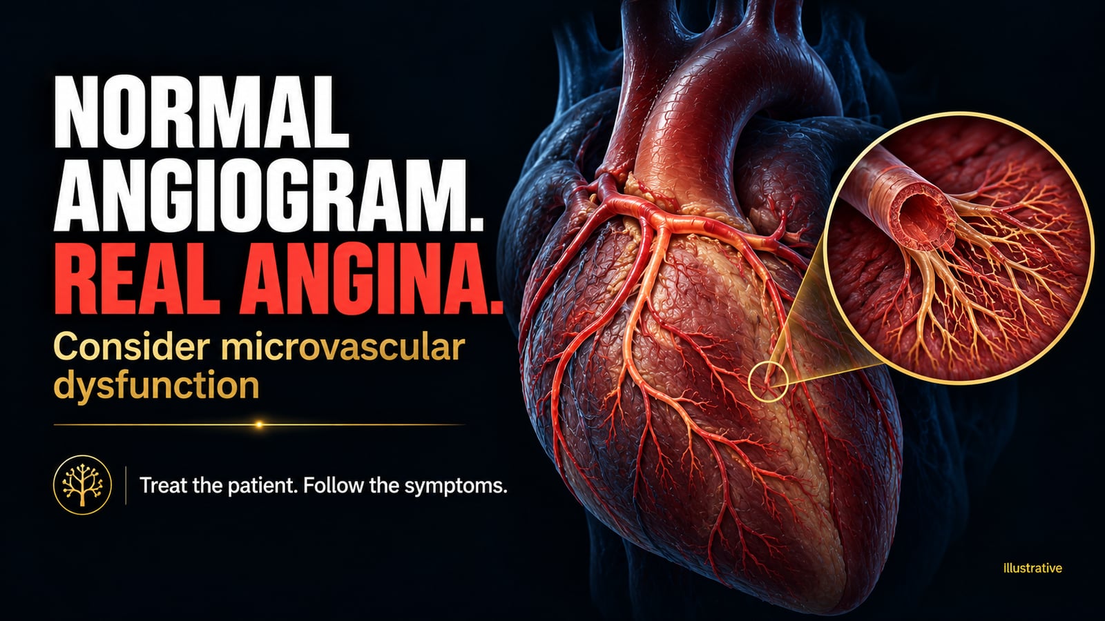
A normal coronary angiogram can be reassuring. It can also leave the reason for a patient's angina unresolved.
That is the clinical tension in this case: a patient with anginal symptoms, and coronary angiography showing no obstructive disease. The images answer an important anatomical question. They do not, by themselves, explain the symptoms.
Angiography shows the lumen of the large epicardial coronary arteries. It does not directly show how the small resistance vessels within the myocardium regulate blood flow. Those vessels must dilate when myocardial oxygen demand rises. If that response is impaired, or microvascular resistance remains abnormally high, the myocardium may receive inadequate flow despite an open epicardial circulation. Spasm can also occur in the epicardial arteries or the microvasculature.
This is why coronary microvascular dysfunction belongs in the differential diagnosis. It is also why a normal angiogram is not sufficient to diagnose it. Angina with non-obstructive coronary arteries describes a clinical presentation; the term ischaemia with non-obstructive coronary arteries requires evidence of myocardial ischaemia. Neither label identifies the underlying mechanism on its own.
The next step starts with the patient again: the circumstances of the discomfort, its duration, associated breathlessness, functional limitation and response to previous treatment. Other cardiac and non-cardiac causes still need consideration. A reassuring angiogram should make our assessment more focused, not make the symptoms less credible.
When symptoms persist despite appropriate initial medical treatment, further testing can help identify what the angiogram cannot. Depending on the clinical question, patient preferences and local expertise, positron emission tomography can assess myocardial blood flow and flow reserve, while stress perfusion cardiac magnetic resonance can help assess myocardial perfusion. Invasive coronary function testing can assess coronary flow reserve and microvascular resistance; acetylcholine testing can identify epicardial or microvascular spasm. The purpose is to connect a treatable mechanism to the patient's symptoms.
That distinction matters because there is no single prescription for every patient with a normal angiogram.
For microvascular dysfunction characterised by reduced flow reserve or increased resistance, without a predominant spasm mechanism, a beta blocker may help by reducing heart rate and myocardial oxygen demand. Calcium-channel blockers or ranolazine may be useful in selected patients, according to the physiological findings, blood pressure, heart rate and response. Benefit varies; response needs reassessment.
When vasospasm is the dominant mechanism, calcium-channel blockers become central. Nitrates can help epicardial spasm, while long-acting nitrates are often less effective in microvascular angina and may be poorly tolerated. Beta blockers are generally avoided in vasospastic angina. Recognising the mechanism can therefore change the choice of drug, not simply its dose.
Risk management remains part of treatment. Blood pressure, lipids, diabetes, smoking and physical activity should be addressed in an individual plan. Statins and an angiotensin-converting enzyme inhibitor or angiotensin receptor blocker may be appropriate according to cardiovascular risk and comorbidities. Antiplatelet treatment is not automatic simply because angina is present. There is no obstructive lesion to stent in this angiogram.
And then we need to ask whether the treatment is helping. Are episodes less frequent? Can the patient walk farther, climb stairs or work with fewer symptoms? Are adverse effects creating a new limitation? Those are clinical outcomes, not secondary details. In the CorMicA trial, treatment guided by coronary function testing improved angina and quality of life at one year. That does not establish fewer myocardial infarctions or longer survival.
Laboratory results, echocardiograms and angiographic images remain essential. They inform diagnosis, prognosis and prevention — including when symptoms are absent. But they acquire their clinical meaning in the context of the person in front of us.
We do not treat an angiographic frame. We treat a patient. A normal image should refine that responsibility, not end it.